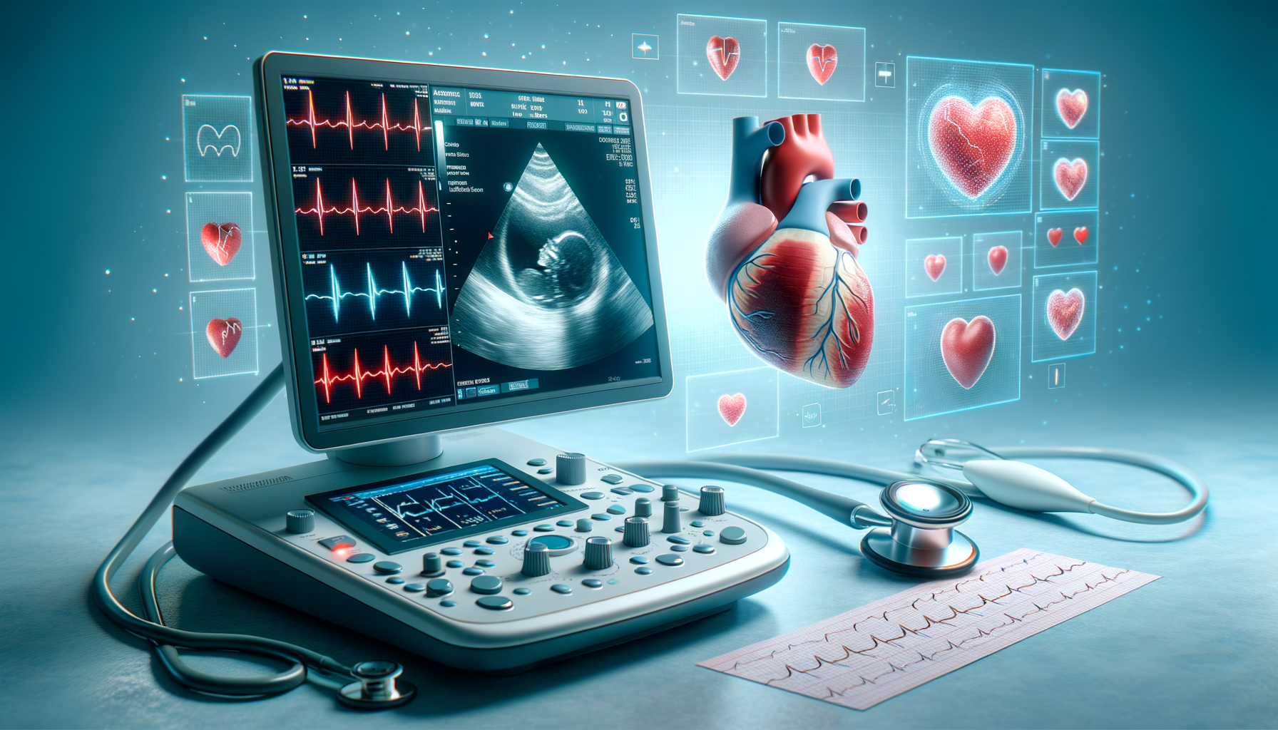
Understanding the Basics of an Echocardiogram
An echocardiogram, often referred to as an “echo,” is a non-invasive diagnostic tool that uses sound waves to produce images of the heart. The procedure is similar to an ultrasound and allows healthcare professionals to observe the heart in real time. This technology provides a detailed look at the heart’s structure and function, making it invaluable in detecting heart diseases and conditions.
The echocardiogram works by sending high-frequency sound waves through a device called a transducer. These sound waves bounce off the heart’s structures and are captured back by the transducer, which then converts them into images. These images are displayed on a monitor, allowing doctors to assess the heart’s chambers, valves, walls, and blood vessels.
There are several types of echocardiograms, including transthoracic echocardiogram (TTE), transesophageal echocardiogram (TEE), stress echocardiogram, and Doppler echocardiogram. Each type serves a specific purpose and is chosen based on the patient’s condition and the information needed. For instance, a TEE is often used when clearer images are required, as it involves inserting the transducer into the esophagus to get closer to the heart.
The Role of Echocardiograms in Diagnosing Heart Conditions
Echocardiograms play a crucial role in diagnosing a wide range of heart conditions. They provide essential information about the heart’s size, shape, and movement, enabling doctors to detect abnormalities such as heart valve issues, congenital heart defects, and cardiomyopathy.
One of the significant advantages of echocardiograms is their ability to assess heart valve function. Valves are vital in directing blood flow through the heart, and any dysfunction can lead to serious health issues. An echocardiogram can identify problems like stenosis (narrowing of the valve) or regurgitation (leaking valve), allowing for timely intervention.
Additionally, echocardiograms are instrumental in diagnosing heart failure. By measuring the ejection fraction, which is the percentage of blood pumped out of the heart’s left ventricle with each beat, doctors can determine how well the heart is functioning. A low ejection fraction is a key indicator of heart failure, prompting further evaluation and treatment.
Comparing Echocardiograms with Other Diagnostic Tools
When it comes to diagnosing heart conditions, echocardiograms are often compared with other diagnostic tools such as electrocardiograms (ECG), cardiac MRI, and CT scans. Each method has its strengths and limitations, and the choice depends on the specific needs of the patient.
Unlike an echocardiogram, an ECG records the electrical activity of the heart and is useful for detecting arrhythmias and heart attacks. However, it does not provide images of the heart’s structure. On the other hand, cardiac MRI and CT scans offer detailed images of the heart and blood vessels but involve exposure to radiation or require the use of contrast agents.
The non-invasive nature and absence of radiation make echocardiograms a preferred choice for many patients. They are particularly advantageous for monitoring heart conditions over time, as repeated exposure to radiation is not a concern. However, in cases where more detailed images are necessary, MRI or CT scans may be recommended.
Preparing for an Echocardiogram: What to Expect
For those scheduled to undergo an echocardiogram, understanding the preparation and procedure can help alleviate any anxiety. Generally, no special preparation is required for a standard transthoracic echocardiogram. Patients can eat and drink as usual, and the procedure is usually performed on an outpatient basis.
During the exam, patients will lie on an examination table, and a technician will apply a special gel to the chest. This gel helps the transducer make secure contact with the skin and improves the transmission of sound waves. The technician will then move the transducer across the chest to capture images of the heart, which are displayed on a monitor.
For a transesophageal echocardiogram, patients may be asked to fast for a few hours before the procedure. This type of echo involves inserting the transducer into the esophagus, so sedation is typically administered to ensure comfort. Patients should arrange for transportation home afterward, as the sedative can impair their ability to drive.
Interpreting Echocardiogram Results: What Do They Mean?
After an echocardiogram, a cardiologist will analyze the images and provide a detailed report of the findings. Understanding these results can offer valuable insights into one’s heart health and guide future medical decisions.
The report will typically include information about the heart’s size and structure, the functioning of the heart valves, and the blood flow through the heart. If any abnormalities are detected, such as thickened heart walls or valve malfunctions, further tests or treatments may be recommended.
It’s important to discuss the results with a healthcare provider to understand their implications fully. While some findings may be benign and require no intervention, others might indicate a need for medication, lifestyle changes, or even surgical procedures. Regular follow-ups and echocardiograms may be necessary to monitor any changes in heart health.

Leave a Reply