Understanding the Echocardiogram: A Window to the Heart
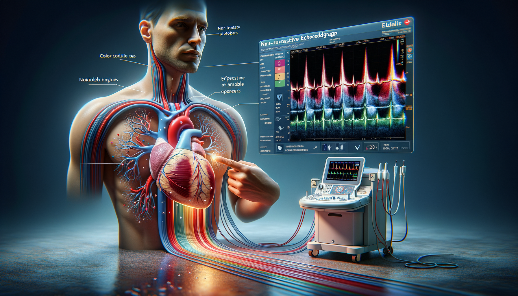
The echocardiogram, often referred to as an “echo,” is a fundamental tool in cardiology that provides a detailed view of the heart’s structure and function. This non-invasive test uses sound waves to create live images of the heart, known as echocardiograms. It is an essential diagnostic tool that helps healthcare professionals assess heart conditions and guide treatment plans. By understanding how an echocardiogram works, patients can appreciate its role in maintaining heart health.
During an echocardiogram, a transducer is placed on the chest, which emits sound waves that bounce off the heart structures. These waves are then captured and translated into images by a computer. The procedure is painless and typically takes about 30 to 60 minutes. The images provide valuable information about the size and shape of the heart, the functioning of its chambers and valves, and the presence of any fluid around the heart.
One of the key benefits of an echocardiogram is its ability to detect abnormalities in the heart’s function and structure. It can identify conditions such as heart valve disease, congenital heart defects, and cardiomyopathy. Moreover, it helps in monitoring the heart’s response to various treatments and in planning surgical interventions if necessary.
Types of Echocardiograms: Tailoring the Test to the Patient’s Needs
There are several types of echocardiograms, each tailored to provide specific information about the heart’s health. The most common type is the transthoracic echocardiogram (TTE), which is performed by placing the transducer on the chest. This standard echo provides a comprehensive overview of the heart’s anatomy and function.
For more detailed images, a transesophageal echocardiogram (TEE) might be recommended. In this procedure, the transducer is passed down the esophagus, providing a closer view of the heart’s structures, particularly useful in evaluating the heart valves and detecting blood clots. While TEE is more invasive than TTE, it offers superior image quality in certain cases.
Stress echocardiograms are another variant, conducted while the heart is under stress, either through exercise or medication. This test helps assess how the heart performs under physical strain and can be crucial in diagnosing coronary artery disease. Additionally, there are specialized echocardiograms like Doppler echocardiograms, which measure the speed and direction of blood flow through the heart, offering insights into the severity of valve problems.
Choosing the right type of echocardiogram depends on the symptoms presented and the specific information needed by the physician, ensuring that the test is both efficient and effective in diagnosing heart conditions.
The Role of Echocardiograms in Diagnosing Heart Conditions
Echocardiograms play a pivotal role in diagnosing a wide range of heart conditions. They provide detailed information that can reveal issues not detectable through other diagnostic methods. By offering a real-time view of the heart in motion, echocardiograms can identify structural abnormalities, functional problems, and the presence of fluid around the heart.
One of the most significant uses of echocardiograms is in diagnosing heart valve diseases. The test can show how well the valves are opening and closing, and detect any abnormalities in blood flow. This information is crucial for determining the severity of valve disorders and planning appropriate treatments.
Congenital heart defects, which are present at birth, can also be identified through echocardiograms. These defects might include holes in the heart, abnormal connections between heart chambers, or improperly formed heart valves. Early detection through echocardiograms allows for timely interventions, which can be life-saving.
In addition to structural issues, echocardiograms can detect cardiomyopathy, a disease of the heart muscle that affects its ability to pump blood effectively. By assessing the thickness and movement of the heart walls, healthcare professionals can diagnose and manage this condition, potentially preventing heart failure.
Overall, echocardiograms are invaluable in the early detection and management of heart conditions, enabling healthcare providers to offer targeted treatments and improve patient outcomes.
Echocardiograms: Differences in Procedure for Women
While the basic principles of echocardiograms remain the same for all patients, there are subtle differences in how the procedure is conducted for women. These differences are primarily due to anatomical variations and the need for tailored approaches to ensure accurate results.
One of the main considerations is breast tissue, which can sometimes obscure the images obtained during a transthoracic echocardiogram. To address this, technicians may need to adjust the position of the transducer or ask the patient to change positions to obtain clearer images. This ensures that the heart structures are visible and that the test results are reliable.
Moreover, women may experience different symptoms of heart disease compared to men, which can influence the focus of the echocardiogram. For instance, women are more likely to present with atypical symptoms of coronary artery disease, such as fatigue and shortness of breath, rather than the classic chest pain. As a result, echocardiograms for women might be tailored to investigate these specific symptoms more thoroughly.
It is also important to consider hormonal factors that can affect heart health in women, such as pregnancy and menopause. During pregnancy, an echocardiogram can be used to monitor heart function, as the heart works harder to support both the mother and the developing fetus. In post-menopausal women, echocardiograms can help assess changes in heart health related to reduced estrogen levels.
Understanding these differences ensures that echocardiograms are conducted effectively for women, providing accurate diagnoses and guiding appropriate treatments.
Conclusion: The Vital Role of Echocardiograms in Heart Health
In conclusion, echocardiograms are an indispensable tool in the realm of cardiology, providing critical insights into the heart’s structure and function. Their non-invasive nature, combined with the ability to offer real-time images, makes them an essential part of diagnosing and managing heart conditions.
By offering tailored approaches to suit individual patient needs, whether through different types of echocardiograms or considerations for gender-specific factors, these tests ensure that healthcare providers can make informed decisions about treatment plans. The early detection of heart conditions through echocardiograms can significantly improve patient outcomes, highlighting the importance of this diagnostic tool in maintaining heart health.
As technology advances, the capabilities of echocardiograms continue to expand, promising even more precise and comprehensive assessments of heart health in the future.

Leave a Reply